3D cell culture


3D IN A FAST AND EASY 2D WORKFLOW

3D Zellkultur Technologien

Warum 3D Zellkultur?

In preclinical drug discovery validation processes, monolayer cell cultures are still predominant. Nevertheless, 2D cultures can only mimic the conditions of physiological tissue to a limited extent, whereas cells in vivo are able to interact in a three-dimensional network. Therefore, results generated from 2D cultures may often be of limited relevance for clinical effectiveness and may contribute to high attrition rates in the drug development process. The employment of spheroid cultures is regarded as a better rational to develop more predictive in-vitro screening assays for preclinical drug development, especially in cancer research.

In spheroid cultures, cells grow in a three-dimensional system with zones of cellular heterogeneity and nutrient and oxygen gradients, to more closely reflect the in-vivo tumor microenvironment. Comparisons of spheroid cultures and 2D monolayer cultures showed functional differences in tumor cell lines, e.g. alterations in protein expression, phosphorylation patterns and responsiveness to inhibitor molecules.

Magnetische 3D Zellkulturtechnik

Die Technologie der magnetischen 3D Zellkultur von Greiner Bio-One beruht auf der Magnetisierung von Zellen mittels biokompatiblem NanoShuttle-PL. Die reproduzierbare Bildung eines Sphäroids pro Well in einer F-Bodenplatte mit zellabweisender Oberfläche wird durch den Einsatz von Magnete entweder durch Levitation oder Bioprinting erreicht, um strukturell und biologisch repräsentative 3D-Modelle in-vitro zu bilden.

Bioprinting 96, 384 & 1536 well

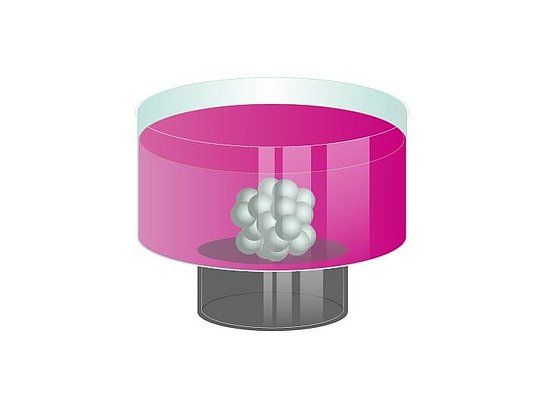
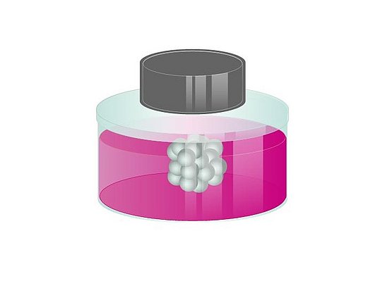
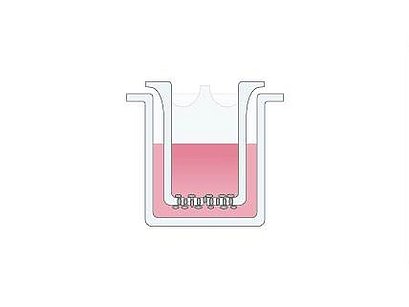
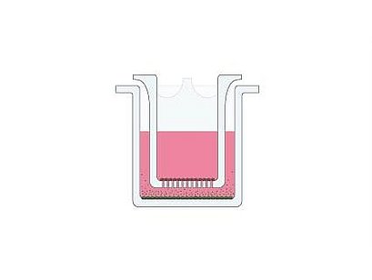
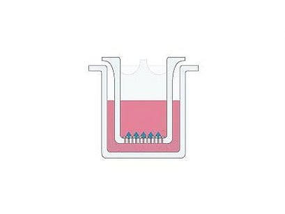
Beim Bioprinting werden magnetisierte Zellen mit Hilfe von schwachen magnetischen Kräften am Näpfchenboden zu Sphäroiden zusammengeführt. Ein Magnet unter jedem Well induziert die Zellaggregation und bildet einen Sphäroid pro Well innerhalb von 15 Minuten bis einigen Stunden.

Levitation 6 & 24 well

Bei der Levitation werden magnetisierten Zellen von einem Magneten, der über dem Zellkulturgefäß platziert wird, in den Schwebezustand angehoben. Die Magnetkräfte wirken hierbei wie ein unsichtbares „Gerüst“, wodurch die Zellen schnell und schonend aggregieren und die Bildung von Zell-Zell-Interaktionen sowie die Expression von ECM-Proteinen induziert wird.

Bio Assay 96 & 384 well

Basierend auf dem Bioprinting werden magnetisierten Zellen zu Sphäroiden oder Ringstrukturen zusammengeführt. Unmittelbar danach schrumpfen/schließen sich diese Strukturen in Abhängigkeit von Zellmigration, Lebensfähigkeit, Zell-Zell-Interaktion und/oder Proliferation.

Die Vorteile der magnetischen 3D Zellkultur (m3D) Technologie:

  • 3D in einem 2D Arbeitsablauf
  • Reproduzierbare Sphäroidbildung
  • Skalierbar – 6 Well bis 1536 Well
  • Durchführung auf einer ebenen Well Oberfläche, optimal für hochauflösende Mikroskopie und HTS
  • Schnelle 3D-Kulturbildung innerhalb von 24 Stunden für die meisten Zelltypen
  • Keine spezielle Ausrüstung oder Medien nötig
  • Einfacher Medienwechsel und Co-Kultivierung verschiedener Zelltypen
  • Kompatibel mit Fluoreszenzmikroskopie, Western Blotting, qRT-PCR, Durchflusszytometrie, Viabilitäts-Assays, Chemilumineszenz, etc.
  • Kompatible mit Automatisierungssystemen
  • Biokompatible Nanopartikel zur Magnetisierung von Zellen

MAGNETISATION WITH NANOSHUTTLE-PL

NanoShuttle-PL is a nanoparticle assembly (~50 nm) consisting of biocompatible components: gold, iron oxide, and Poly-L-Lysine (PLL). Although NanoShuttle-PL is not itself an FDA-approved product for use in humans, the constituent components are themselves biocompatible. The cells are magnetised by electrostatically attaching small amounts of NanoShuttle-PL non-specifically to cell membranes via PLL at a concentration of around 50 pg/cell. Magnetised cells will appear peppered with dark nanoparticles after incubation, giving a speckled appearance. A small magnetic force of 30 pN/cell is enough to levitate and assemble cells without causing any harm.

NANOSHUTTLE-PL - THE BIOCOMPATIBILITY

  • Will not affect proliferation, viability, metabolism, inflammatory or oxidative stress, phenotype and/ or other cell functions
  • Does not bind any specific receptors, works with all cell types
  • Will release from the cell over 7-8 days into the surrounding extracellular matrix
  • Does not cause any chromosomal abnormalities in cells and does not lead to genomic instability


Im Gegensatz zur magnetischen Levitation werden beim magnetischen 3D-Bioprinting magnetisierte Zellen mit Hilfe von schwachen magnetischen Kräften am Näpfchenboden zu Sphäroiden zusammengeführt. Ein Magnet unter jedem Well induziert die Zellaggregation und bildet einen Sphäroid pro Well innerhalb von 15 Minuten bis einigen Stunden. Danach können die Sphäroide ohne Magnetkraft langfristig kultiviert werden. Dieses System überwindet die Grenzen anderer Plattformen, indem es eine schnelle Bildung von Sphäroiden ermöglicht, die Größe der Sphäroide in Abhängigkeit von der Zellzahl reproduzierbar und skalierbar ist. Die Technologie ist für Hochdurchsatz-Anwendungen (96, 384 und 1536 Well) geeignet und nicht auf spezifische Zelltypen beschränkt. 

Das 3D-Bioprinting in Verbindung mit handelsüblichen standardisierten biochemischen Testverfahren zur kontinuierlichen Beurteilung der Zell-Viabilität und anderer Funktionen bietet eine ideale Kombination für das Compound Screening im Hochdurchsatz dar.

Durch die Nutzung magnetischer Kräfte zum Festhalten der magnetisierten Sphäroide während der Aspiration wird das Hinzufügen und Entfernen von Lösungen erleichtert und der Sphäroidverlust begrenzt. Sphäroide können durch den Einsatz von Magnetwerkzeugen, wie dem MagPen aufgenommen und zwischen den Näpfchen übertragen werden. Darüber hinaus können die magnetischen Kräfte auch genutzt werden, um Co-Kulturen mit feiner räumlicher Anordnung zu schaffen.

Die magnetische Levitation ist eine einfache Methode, um native Gewebeumgebungen in vitro herzustellen. Die magnetisierten Zellen werden von einem Magneten, der über dem Zellkulturgefäß platziert wird, in den Schwebezustand angehoben. Die Magnetkräfte wirken hierbei wie ein unsichtbares „Gerüst“, durch dessen Hilfe die Zellen schnell und schonend aggregieren und die Bildung von Zell-Zell-Interaktionen, sowie die Expression von ECM-Proteinen induziert. Die 3D-Zellkulltur Bildung erfolgt ohne Zugabe von künstlichen Substraten oder der Verwendung von speziellen Medien oder Geräte und kann langfristig kultiviert werden. Durch die sanfte Methode der magnetischen Levitation können die Kulturen eine Morphologie im Makroskala bilden, welche ihrem nativen Gewebe am nächsten kommt.

Die Hauptanwendung dieser Technologie ist die Herstellung von 3D-Zellkulturen unter verschiedenen Kultivierungsbedingungen, um sie mit gängigen biologischen Techniken, wie immunhistochemischen Analysemethoden und Western Blots, zu untersuchen.

Die aktuellen Standards für das Wirkstoffscreening sind Tiermodelle; sie repräsentieren auf der einen Seite menschliches Gewebe von Interesse sind aber auf der anderen Seite teuer, knapp und mit ethischen Herausforderungen verbunden. 2D In-vitro-Assays imitieren schlecht die native zelluläre Umgebung und damit die menschliche in vivo-Reaktion, bieten aber einfache Hochdurchsatztests. Es besteht ein Bedarf an In-vitro-Assays, welche sowohl die menschliche in vivo Reaktion als auch den Hochdurchsatz kombinieren.

Aus diesem Grund haben wir einen Viabilitäts-Assay, den BiO-Assay, entwickelt. Basierend auf dem magnetischen 3D-Bioprinting werden die mit NanoShuttle-PL (NS) magnetisierten Zellen zu Sphäroide oder Ringstrukturen zusammengeführt.

Unmittelbar im Anschluss schrumpfen/schließen sich diese Strukturen in Abhängigkeit von Zellmigration, Lebensfähigkeit, Zell-Zell-Interaktion und/oder Proliferation und variieren je nach Wirkstoff-Dosierung. Die Kontraktion / der Ringschluss der Kultur ist in der Regel innerhalb von 24 Stunden abgeschlossen und die Bilder werden im Batch-Verfahren verarbeitet, um schnell Toxizitätsdaten zu erhalten. Da der Assay markierungsfrei ist, stehen die verbleibenden Ringe oder Sphäroide für weitere Experimente zur Verfügung (IHC, Western Blot, Genomik, etc.).

Der BiO-Assay kann verwendet werden, um die Kontraktion von Ringen und Sphäroiden zu verfolgen, welche verschiedene Situationen darstellen kann. Bei Ringen kann das Schließen des Rings eine Wundheilung abbilden, wobei Zellen daran arbeiten, den Hohlraum in der Mitte des Rings zu schließen. Darüber hinaus können Ringe ähnlich geformte Gewebe wie Blutgefäße abbilden, in denen Dilatation und Kontraktion untersucht werden kann. Bei Sphäroiden steht die Kontraktion im Zusammenhang mit der sphäroidischen Aussbildung, wobei der Test makroskopisch misst, wie gut die Zellen interagieren und migrieren, um eine kompetente Struktur aufzubauen.

Der BiO-Assay kombiniert 3D-Zellkultur mit Hochdurchsatz- und Hochauflösenden-Tests, um die in vivo-Reaktion in vitro effektiv vorherzusagen.

MagPen - Your smart assistant for 3D cell culture transfer

The MagPen facilitates easy and fast transfer and collection of magnetised cell cultures without disrupting their microtissue architecture. Cells, magnetised and cultured by Magnetic 3D Cell Culture (M3D), can be transposed by a simple “pick up-and-drop”-step. Additionally to that, the MagPen can be used to create and organize co-cultures by combining different magnetised 3D cell cultures.
The MagPen is availible as single version and as Multi-MagPen in 24 and 96 well format for simultaneous transfer of various cell cultures in one step.

MagPen - The principle

  • Fast, easy and simultaneous transfer of multiple 3D cell cultures without pipetting
  • Complete media change by a simple “pick up-and-drop”- step
  • Simplified co-culturing of different cell types
  • Ideal for easy immunohistochemistry staining, blocking, and washing of spheroids

ThinCert® Zellkultureinsätze

Die ThinCert®-Zellkultureinsätze eignen sich für ein breites Spektrum von Anwendungen, darunter:

  • Transport-, Sekretions- und Diffusionsstudien
  • Migrationsexperimente
  • Zytotoxizitätstests
  • Co-Kulturen
  • transepitheliale elektrische Widerstandsmessungen (TEER) 
  • Primärzellkulturen

Untersuchung von Migration und Invasion

Die Zellmigration spielt eine wichtige Rolle bei physiologischen und pathologischen Prozessen während der Embryonalentwicklung. Sowie der Wundheilung, der Immunreaktion, bei Entzündungen und der Tumorentstehung. Der Filter-Assay ist ein Standard-In-vitro-Modell zur Untersuchung der Zellmigration. Dieser wird in Zellkultureinsätzen mit 8,0 μm großen Poren der ThinCert®-Membran durchgeführt. Die Zellmigration wird hierbei aus dem oberen Kompartiment in Richtung einer Chemo-Attraktionsquelle im unteren Kompartiment unterstützt und beinhaltet.

Co-Kultur

Bei der Co-Kultur werden die Interaktionen zwischen Immunzellen untersucht. Darüber hinaus umfasst sie weitere vielfältige Anwendungen wie die Stimulierung der Zellproliferation, die Aufrechterhaltung der Zelldifferenzierung und die Wiederherstellung heterozellulärer Funktionen in vitro (z. B. Blut-Hirn-Schranke). Bei den ThinCert®Zellkultureinsätzen können die Zellen im oberen und unteren Kompartiment angesiedelt werden. Die 0,4 µm oder 1,0 µm großen Poren der ThinCert®Membran ermöglichen den Austausch von Molekülen zwischen den beiden Zellpopulationen.

Transport-Studien

Transportstudien gehören zu den häufigsten Anwendungen von Zellkultureinsätzen. Ziel ist es, aus einzelnen Zellen ein funktionsfähiges Epithel zu rekonstruieren. Außerdem soll ein aktiver Transport von Substanzen von einem Kompartiment durch das Epithel in das andere Kompartiment untersucht werden. ThinCert® Zellkultureinsätze mit 0,4 µm Poren und transluzenten Membranen werden für Transportuntersuchungen empfohlen.

Organotypische und Air-Lift-Kultur

In der organotypischen Kultur kann ein Gewebe über einen längeren Zeitraum am Leben erhalten werden. Bei der Geweberekonstruktion hingegen wird das Gewebe de novo aus einzelnen Zellen erzeugt. Bei beiden Verfahren werden Zellkultureinsätze mit 0,4 bis 3,0 µm Poren verwendet. Das Gewebe wächst in vitro an der Luft-Flüssigkeits-Grenzfläche ohne Einschränkungen durch den Gasaustausch. Darüber hinaus dient bei einigen Gewebetypen die direkte Exposition der kultivierten Zellen gegenüber der umgebenden Atmosphäre als unverzichtbarer Differenzierungsstimulus.

  • Hängende Geometrie
  • Verbesserte Zelladhäsion durch physikalische Oberflächenbehandlung
  • Vereinfachtes Pipettieren durch Self-Lift-Geometrie
  • Verbesserter Pipettierzugang und Gasaustausch

Für licht- oder elektronenmikroskopische Untersuchungen können die Membranen einfach mit Hilfe eines Skalpells vom Gehäuse gelöst werden. Auch im abgelösten Zustand verbleibt die Membran flach, ohne sich aufzurollen, was weitere Arbeitsschritte wesentlich vereinfacht. Aufgrund ihrer hohen chemischen Widerstandsfähigkeit gegenüber verschiedenen Lösungsmitteln ist die Membran für zahlreiche Fixierprotokolle für Zellen geeignet. Durch ihre spezielle Aufhängung halten ThinCert® Zellkultureinsätze jederzeit einen Abstand zum Wellboden, wodurch dort kultivierte Zellen stets vor Beschädigung geschützt sind. Zu den Seitenwänden ist mit Hilfe von Distanzhaltern immer ein sicherer Mindestabstand eingehalten, der einen Kapillarsog zwischen Wellinnenwand und Außenwand des Einsatzes verhindert. D.h. der Stoffaustausch zwischen Well und ThinCert® erfolgt ausschließlich über die poröse Membran. ThinCert® Zellkultureinsätze sitzen exzentrisch in den Wells und weichen beim Einführen einer Pipette nach oben aus. Wenn die Pipette wieder entfernt wird, nehmen die ThinCert® ihre ursprüngliche Position automatisch wieder ein. Diese Eigenschaft wird Self-Lift-Geometrie genannt.

  • Kleine Porengrößen (Durchmesser 0,4 und 1 μm) sind sowohl für Co-Kultivierungen als auch für Transport-, Sekretions- und Diffusionsstudien kleiner Moleküle geeignet.
  • Größere Porengrößen (Durchmesser 3 und 8 μm) eignen sich für Migrations- und Invasionsstudien.
  • Transparente Membranen (allgemein: Membranen mit niedriger Porendichte) sind sowohl für die Licht- als auch für die Elektromikroskopie einsetzbar.
  • Transluzente Membranen (allgemein: Membranen mit hoher Porendichte) sind sowohl für die Elektronenmikroskopie als auch für TEER und Transportassays einsetzbar.

Material:

ThinCert® Zellkultureinsätze sind aus einem hochwertigen transparenten Polystyrol-Gehäuse mit Böden aus Polyethylenterephthalat (PET) Kapillarporenmembranen gefertigt. Beide Materialien sind USP Class VI zertifiziert und absolut zellverträglich. Das Gehäuse und die Membran sind mit Hilfe eines automatisierten Siegelprozesses miteinander verbunden, was zu einer hochfesten und –dichten Verbindung führt, ohne die Membran zu schwächen. Die physikalische Oberflächenbehandlung der PET-Membran ermöglicht eine optimale Adhärenz und Wachstumscharakteristik kultivierter Zellen. Alle Kapillarporen in einer Membran weisen einen sehr gleichmäßigen Durchmesser auf. Diese Einheitlichkeit gewährleistet zuverlässige und gleichbleibende Austauschraten zwischen den beiden Kompartimenten und sorgt somit für Reproduzierbarkeit bei der Durchführung mehrerer Experimente.

Kits:

Als gebrauchsfertige Kits enthalten die Packungen bereits CELLSTAR® Multiwell-Platten in entsprechender Anzahl. Der vollautomatische Herstellungsprozess beinhaltet eine automatisierte doppelte optische Kontrolle jedes einzelnen hergestellten Einsatzes. Eine abschließende Bestrahlung stellt die Sterilität der individuell in Blister verpackten Zellkultureinsätze und Multiwell-Platten sicher.

CELLSTAR® Cell-Repellent Surface

Im Gegensatz zu Standard-Gewebekulturoberflächen, die optimiert sind, um die Bedingungen für die Zellbindung zu verbessern, wurde die zellabweisende Oberfläche entwickelt, um die Zellbindung effektiv zu unterbinden. CELLSTAR® Zellkulturgefäße mit einer zellabweisenden Oberfläche verhindern zuverlässig die Zellbindung in Suspensionskulturen von semi-adhärenten und adhärenten Zelllinien, bei denen die für die Suspensionskultur üblichen hydrophoben Standardoberflächen unzureichend sind.

Für die Bildung von Sphäroiden, Stammzellaggregaten und selbstorganisierten runden Clustern, die als 3D-Zellkulturmodelle verwendet werden, muss die Zell-Zell-Interaktion über die Interaktion zwischen den Zellen und der Zellkulturgefäßoberfläche dominieren. 

CELLSTAR® Zellkulturgefäße mit zellabweisender Oberfläche verhindern wirkungsvoll die Zellanhaftung und fördern die spontane Bildung dreidimensionaler Sphäroide durch Gravitation: ein einzelner Sphäroid pro Well in Rundboden-Mikroplatten oder mehrere Sphäroide in Flachbodenplatten, Schalen und Flaschen.

Langfristige Inkubationen von Hydrogelkulturen werden häufig als Ansatz zur Nachahmung einer 3D-Umgebung durchgeführt. Wenn bei diesem Ansatz Standard-Gewebekulturgefäße verwendet werden, neigen einige Zellen dazu, aus dem Hydrogel zu migrieren und eine 2D-Subkultur auf der Gefäßoberfläche zu bilden. Die Analyse einer solchen Zellpopulation führt daher zu gemischten Daten aus 2D- und 3D-Zellkulturen. Durch den Einsatz von CELLSTAR® Zellkulturgefäße mit einer zellabweisenden Oberfläche für Hydrogelkulturen wird die Bildung von 2D-Subkulturen effektiv unterdrückt.

Applikationen & Zelllinien

Welche Plattform ist die beste für Ihre Applikation?

Stem Cell Lines

Cancer Cell Lines

Primary Cell Lines

Other Cell Lines

Pre-adipocyte stem cell

KPC pancreatic ductal adenocarcinoma


Pancreatic ß-cells (EndoC-ßH3)

Neural stem cells

LN229 – Glioblastoma cells

Primary Glioblastoma cells

Human Astrocyte

Neural crest-derived mesenchymal stem cell

A549 — Lung epithelial adecarcenoma

Valvular interstitial cells (VICs)

Bend (brain endothelial)

Mesenchymal stem cell

HepG2 — Human liver carcinoma

Human lung primary cells:

  • epithelial
  • endothelial
  • fibroblasts
  • smooth muscle

3T3 fibroblasts

Dental pulp stem cell

PC3 — Human prostate cancer

Valvular endothelial cells (VECs)

Adipocyte


H-4-II-E — Rat hepatoma, liver

Aortic valve co-cultures (AVCCs)

Huvec - Human umbilical endothelial cells


MDA-231 - Human breast cancer

Primary mouse heart cells

HPF - Human pulmonary fibroblast


LNCaP - Prostate cancer cell line

Human Primary vascular smooth muscle

SMC - Tracheal smooth muscle cell


Ovarian cancer cells

Primary Miomytrial Smooth muscle

HEK293 - Human embryonic kidney


Panc-1 - Pancreatic cancer cell

Primary human hepatocytes

MCF-10A - Breast epithelial cell line


Cancer associated fibroblasts

Primary pancreatic cancer cells

Fibroblast


Triple negative inflammatory breast cancer

Primary tissue from PDX

Chondrocytes


Caki-1 – Human renal cancer cell line

Primary fibroblasts

T-cells


Osteosarcoma

Keratinocytes

A10 - Rat vascular smooth muscle


HCT116 - colon cancer cell line


DU145


Murine embryonic fibroblasts (iMEF)


LOVO cells


DT66066 cells


Spinal cord cells


Endothelial cell


Peripheral blood mononuclear cells (PBMCs)


Monocytes

Publikationen

Year

Author / Title / Link

Product

Application Area

Cell Type

2022

Chen, G.; Liu, W.; Yan, B.: Breast Cancer MCF-7 Cell Spheroid Culture for Drug Discovery and Development

Cell-Repellent

Breast Cancer

MCF-7 cells (Human Breast Cancer)

2022

Neebe et al. Small integral membrane protein 10 like 1 downregulation enhances differentiation of adipose progenitor cells Biochemical and Biophysical Research Communications

Cell-Repellent

Cancer / Lipoma Formation

Lipoma Cells

2022

Seitlinger et al. Vascularization of Patient-Derived Tumoroid from Non-Small-Cell Lung Cancer and Its Microenvironment

ThinCert®

Cancer

Human Pulmonary Fibroblasts,

Human Umbilical Vein Endothelial Cells,

Human Mesenchymal Stem Cells,

Lung Tumor Cells 

2021

Batista et al. Three-Dimensional Adipocyte Culture as a Model to Study Cachexia-Induced White Adipose Tissue Remodeling

m3D

Cancer Research

Stem Cells

Adipose Tissue,

Sem Cell Adipocyte

2021

Jaromi et al. KRAS and EGFR Mutations Differentially Alter ABC Drug Transporter Expression in Cisplatin-Resistant Non-Small Cell Lung Cancer

m3D

Cancer Resaerch

A549 (KRAS),

PC9 (EGFR)

2020

Kiss et al. Cisplatin treatment induced interleukin 6 and 8 production alters lung adenocarcinoma cell migration in an oncogenic mutation dependent manner  

m3D

Cancer Research

Migration/Invasion Assay

Wound Healing

Primary Lung Cancer,

549 and PC-9 Human Lung Adenocarcinoma

2020

Souza-Araújo et al. Three-Dimensional Cell Culture Based on Magnetic Fields to Assemble Low-Grade Ovarian Carcinoma Cell Aggregates Containing Lymphocytes

m3D

Cancer Research

Patient Cells,

Ovarian Carcinoma,

Lymphocytes

2019

Leenhardt et al. Ultrasound-induced cavitation enhances the efficacy of Chemotherapy in a 3D Model of Pancreatic Ductal Adenocarcinoma with its microenvironment

m3D

Cancer Research

Murine Embryonic Fibroblasts (iMEF),

KPC Pancreatic Ductal Adenocarcinoma (PDA),

T66066 Cells

2019

Mejía-Cruz et al. Generation of Organotypic Multicellular Spheres by Magnetic Levitation: Model for the Study of Human Hematopoietic Stem Cells Microenvironment

m3D

Cancer Research

Stem Cells

Human Hematopoietic Stem Cells,

Human Bone Marrow-Mesenchymal Stem Cells,

Umbilical Cord Blood-Hematopoietic Stem Cells

2018

Degadi et al. Klotho inhibits EGF-induced cell migration in Caki-1 cells through inactivation of EGFR and p38 MAPK signaling pathways

m3D

Cancer Research

Wound Healing

Caki-1

2018

Eckhardt et al. Clinically relevant inflammatory breast cancer patient-derived xenograft-derived ex vivo model for evaluation of tumor-specific therapies

m3D

Cancer Research

Screening / Toxicity

Triple Negative Inflamatory Breast Cancer

2018

Hou et al. Advanced Development of Primary Pancreatic Organic Tumor Models for High-Throughput Phenotypic Drug Screening

m3D

Cancer Research

Screening / Toxicity

Triple Negative Inflamatory Breast Cancer

2017

Noel et al. Preparation and Metabolic Assay of 3-dimensional Spheroid Co-cultures of Pancreatic Cancer Cells and Fibroblasts

m3D

Cancer Research,

Co-Culture

Panc-1 - Pancreatic Cancer Cell

Fibroblast

2016

Hau et al. Dose enhancement and cytotoxicity of gold nanoparticles in colon cancer cells when irradiated with kilo‐ and mega‐voltage radiation

m3D

Cancer Research

Migration/Invasion Assay

LOVO cells,

Colon adenocarcinoma

2016

Pan et al. miR-509-3p is clinically significant and strongly attenuates cellular migration and multi-cellular spheroids in ovarian cancer

m3D

Cancer Research

Wound Healing

Ovarian ccancer cells

2014

Jaganathan et al. Three-dimensional in vitro co-culture model of breast tumor using magnetic levitation

m3D

Cancer Research

Tissue Engineering / Reconstruction

Co-Culture

MDA-231,

Fibroblast

2013

Becker, J. L.; Souza, G. R.: Using space-based investigations to inform cancer research on Earth

m3D

Cancer Research

Glioblastoma

2010

Becker, J. L.; Souza, G. R.: Invasive glioblastoma cells acquire stemness and increased Akt activation

m3D

Cancer Research

Stem Cells

LN229 - Glioblastoma,

Normal Human Astrocyte,

Primary Glioblastoma

2010

Glauco et al. Three-dimensional tissue culture based on magnetic cell levitation

m3D

Cancer Research

Stem Cells

Co-Culture

LN229 - Glioblastoma,

Normal Human Astrocyte,

Neural Stem Cell

Year

Author / Title / Link

Product

Application Area

Cell Type

2022

Baarsma et al. Epithelial 3D-spheroids as a tool to study air pollutant-induced lung pathology

m3D

Screening/Toxicity

Human Bronchial Epithelial (BEAS-2B)

2022

Cromwell et al. Multifunctional profiling of triple-negative breast cancer patient-derived tumoroids for disease modeling

m3D

Screening/Toxicity

Cancer Research

Microfluids

Patient-Derived Tumor Explant,

TU-BcX-4IC,

Breast Cancer,

PDX Organoids (PDX-O) 

2022

Fernandez-Vega et al. Lead Identification using 3D Models of Pancreatic Cancer

m3D

Screening/Toxicity

HTS

Patient Samples

Pancreatic Cancer Cells

2021

Rodboon et al. Development of high-throughput lacrimal gland organoid platforms for drug discovery in dry eye disease

m3D

Screening/Toxicity

Stem Cells

Myoepithelial Cells (MEC)

Lacrimal Gland Ctem Cells

2021

Trindade Caleffi et al. Magnetic 3D cell culture: State of the art and current advances

m3D

Screening/Toxicity

Cardiomyocytes,

Bronchial and Pancreatic Cells

2020

Burnham et al. A Scalable Approach Reveals Functional Responses of iPSC Cardiomyocyte 3D Spheroids

m3D

Screening/Toxicity

Electrophysiology

Cardiomyocytes and Fibroblast

Co-Culture

2019

Baillargeon et al. Automating a Magnetic 3D Spheroid Model Technology for High-Throughput Screening

m3D

Screening/Toxicity

Cancer Research

Pancreatic Cancer Cells

2018

Souza et al. Comparative Assay of 2D and 3D Cell Culture Models: Proliferation, Gene Expression and Anticancer Drug Response

m3D

Screening/Toxicity

Cancer Research

Prostate Cancer Cell Lines: PC-3,

LNCaP and DU145

2017

Desai, P. K.; Tseng, H.; Souza, G. R.: Assembly of hepatocyte spheroids using magnetic 3D cell culture for CYP450 inhibition/induction

m3D

Screening/Toxicity

Primay Human Hepatocytes

2017

Souza et al. Magnetically bioprinted human myometrial 3D cell rings as a model for uterine contractility

m3D

Screening/Toxicity

Primary Miomytrial Smooth Muscle

2016

Tseng et al. A high-throughput in vitro ring assay for vasoactivity using magnetic 3D bioprinting

m3D

Screening/Toxicity

Wound Healing

A10 - Rat Vascular Smooth Muscle,

Human Primary Vascular Smooth Muscle

2015

Tseng et al. A spheroid toxicity assay using magnetic 3D bioprinting and real-time mobile device-based imaging

m3D

Screening/Toxicity

3T3 Fibroblast

2013

Haisler et al. Three-dimensional cell culturing by magnetic levitation

m3D

Screening

Tissue Engineering / Reconstruction

Cancer Research

HepG2 - Human Liver Carcinoma,

A549 - Lung Epithelial Adecarcenoma,

PC3 - Human Prostate Cancer,

LN229 - Glioblastoma,

Huvec - Human Umbelical Endothelial Cells,

H-4-II-E - Rat Hepatoma, Liver,

T3T - Mouse fFbroblast,

HPF - Human Pulmonary Fibroblast,

SMC - Tracheal Smooth Muscle,

MDA-231 - Human Breast Cancer(a),

HEK293 - Human Embrionic Kidney,

MCF-10A - Breast Epithelial Cell Line,

LNCaP - Prostate Cancer Cell Line

Year

Author / Title / Link

Product

Application Area

Cell Type

2022

Baarsma et al. Epithelial 3D-spheroids as a tool to study air pollutant-induced lung pathology

Cell-Repellent

Lung pathology

Human Bronchial Epithelial (BEAS-2B)

2022

Castillo et al. Human Air-Liquid-Interface Organotypic Airway Cultures Express Significantly More ACE2 Receptor Protein and Are More Susceptible to HCoV-NL63 Infection than Monolayer Cultures of Primary Respiratory Epithelial Cells

ThinCert®

COVID

Primary Human Respiratory Epithelial Cells

2022

Chansaenroj et al. Magnetic bioassembly platforms towards the generation of extracellular vesicles from human salivary gland functional organoids for epithelial repair

m3D

Tissue Engineering / Reconstruction

Stem Cells

Human Dental Pulp Stem Cells (hDPSC),

Salivary Gland

2022

Tan, C. T., Leo, Z. Y., Lim, C. Y.: Generation and integration of hair follicle-primed spheroids in bioengineered skin constructs

ThinCert®

Skin research

N/TERT-1 Keratinocytes,

Human Primary Adult Keratinocytes,

Primary Neonatal Keratinocytes,

Human Hair Follicle DP Cells 

2021

Bing, et al. Human organoid biofilm model for assessing antibiofilm activity of novel agents

ThinCert®

Skin Model / Biofilm

N/TERT Keratinocytes

2019

Bowser, D. A.; Moore, M. J.: Biofabrication of neural microphysiological systems using magnetic spheroid bioprinting

m3D

Tissue Engineering / Reconstruction

Spinal Cord Cells

2018

Tseng et al. Three-Dimensional Magnetic Levitation Culture System Simulating White Adipose Tissue

m3D

Tissue Engineering / Reconstruction

Endothelial Cell,

Pre-Adipocyte Stem Cell

2016

Hogan et al. Assembly of a functional 3D primary cardiac construct using magnetic levitation

m3D

Tissue Engineering / Reconstruction

Primary Mouse Heart Cells,

Fibroblast,

Endothelial Cells

2016

Lin et al. Nanoparticle improved stem cell therapy for erectile dysfunction in a rat model of cavernous nerve injury

m3D

Tissue Engineering / Reconstruction

Stem Cells

Mesenchymal Stem Cell

2013

Timm et al. A high-throughput three-dimensional cell migration assay for toxicity screening with mobile device-based macroscopic image analysis

m3D

Wound Healing

Screening/Toxicity

HEK293 - Kidney,

Primary Tracheal Smooth Muscle

2013

Tseng et al. A three-dimensional co-culture model of the aortic valve using magnetic levitation

m3D

Tissue Engineering / Reconstruction 

Co-Culture

Valvular Interstitial Cells (VICs),

Valvular Endothelial Cells (VECs),

Aortic Valve Co-Cultures (AVCCs)

2013

Tseng et al. Assembly of a three-dimensional multitype bronchiole coculture model using magnetic levitation

m3D

Tissue Engineering / Reconstruction 

Primary Human Lung Cells: Epithelial, Endothelial, Fibroblast, Smooth Muscle

Year

Author / Title / Link

Product

Application Area

Cell Type

2021

Avelino et al. Mass spectrometry-based proteomics of 3D cell culture: A useful tool to validate culture of spheroids and organoids

m3D

Stem Cells

Tissue Engineering / Reconstruction

3T3,

Pre-Adipocyte Stem Cells,

Adipocytes

2019

Ferreira et al. A magnetic three‐dimensional levitated primary cell culture system for the development of secretory salivary gland‐like organoids

m3D

Stem Cells

Salivary Gland Derived Cells

2018

Adine et al. Engineering innervated secretory epithelial organoids by magnetic three-dimensional bioprinting for stimulating epithelial growth in salivary glands

m3D

Stem Cells

Tissue Engineering / Reconstruction

Neural Crest-Derived Mesenchymal Stem Cell,

Dental Pulp Stem Cell

2013

Daquinag, A. C., Souza, G. R.; Kolonin, M. G.: Adipose tissue engineering in three-dimensional levitation tissue culture system based on magnetic nanoparticles

m3D

Stem Cells

Tissue Engineering / Reconstruction

Primary Stroma,

3T3,

Bend (Brain Endothelial)  

Year

Author / Title / Link

Product

Application Area

Cell Type

2021

Vu et al. Scaffold-free 3D cell culture of primary skin fibroblasts induces profound changes of the matrisome

m3D

Tissue Engineering / Reconstruction

Primary Skin Fibroblasts

2019

Antonino et al. Three-dimensional levitation culture improves in-vitro growth of secondary follicles in bovine model

m3D

-

Bovine Secondary Follicles,

Oocyte

2019

Klinder et al. Comparison of different cell culture plates for the enrichment of non-adherent human mononuclear cells

Cell-Repellent

-

Peripheral Blood Mononuclear Cells (PBMCs),

Monocytes

2019

Nagaraju et al. Myofibroblast modulation of cardiac myocyte structure and function

ThinCert®

Myocardial Infarction

Cardiomyocytes,

Fibroblasts

2019

Urbanczyk et al. Controlled Heterotypic Pseudo-Islet Assembly of Human b-Cells and Human Umbilical Vein Endothelial Cells Using Magnetic Levitation

m3D

Co-Culture

Tissue Engineering / Reconstruction

Pancreatic ß-Cells (EndoC-ßH3),

Human Umbilical Vein Endothelia Cells (HUVECs)

2013

Castro-Chavez et al. Effect of lyso-phosphatidylcholine and Schnurri-3 on osteogenic transdifferentiation of vascular smooth muscle cells to calcifying vascular cells in 3D culture

m3D

-

Human Umbilical Vein Endothelia Cells (HUVECs)

Downloads & Media

Magentische 3D Zellkulturtechnik - Tools zur Überbrückung der Grenzen zwischen 2D und 3D

Moderator: Gluaco R. Souza

Jahr: 2020, Sprache: Englisch

Lernziele:

  • Grundlagen der magnetischen 3D-Zellkultur und des magnetischen 3D-Bioprinting
  • Die wichtigsten Vorteile der 3D-Zellkultur gegenüber anderen Techniken
  • Breite Anwendungen der 3D-Zellkultur
  • Anwendungen in der Immunbiologie
  • Vergleich zwischen magnetischer 3D-Zellkultur und In-vivo-Ergebnissen in Zusammenarbeit mit dem MD Anderson Cancer Center

Automatisierte Sphäroidproduktion durch magnetische Zellanordnungen für reproduzierbare HTS-Anwendungen

Moderator: Glauco R. Souza (Greiner Bio-One) & Michael Nosswitz (Tecan Schweiz)
Jahr: 2023, Sprache: Englisch

Lernziele:

  • Lernen Sie, wie Sie magnetisches 3D-Bioprinting nutzen können, um Automatisierung und optimale Bildgebungsbedingungen zu ermöglichen
  • Entdecken Sie, wie Sie die Sphäroid-Produktion automatisieren können, um Abweichungen in der Homogenität zu reduzieren
  • Erfahren Sie, wie Sie die Qualitätskontrolle für die Sphäroidbildung und -produktion automatisch überwachen können
  • Erfahren Sie, wie Sie die Medikamentendosierung für Synergieexperimente vereinfachen und automatisieren können.

Year

Title

Conference

2019

Three-dimensional cytotoxicity assay using magnetic 3D bioprinting for measuring CAR T cell function in heterogeneous solid tumor microenvironments

American Association of Cancer Research Annual Meeting, Atlanta, GA

2018

Automated, Image-Based T Cell Mediated Cytotoxicity Assessments using 2D and 3D Target Cell Models

SLAS 2018

2018

3D Cultures of iPSC-derived Human Motor Neurons & Tracheal Smooth Muscle Cells in HTS Format Using Magnetic 3D Bioprinting

Society for Neuroscience Annual Meeting, San Diego, CA

2018

A PDX-DerivedEx-Vivo Tumor Tissue Array Platform Utilizing Magnetic 3D Bioprinting for The Identification of Tumor-Specific Therapies

American Association of Cancer Research Annual Meeting, Chicago, IL

2018

Validation of Magnetic 3D Spheroid Bioprinting in Combination with a BlueWasher

Society of Lab Automation and Screening Annual Conference and Exhibition, San Diego, CA

2016

Development of spheroids derived from tumor biopsies and patient-derived xenografts using magnetic 3D bioprinting

American Association of Cancer Research Annual Meeting, New Orleans, LA

2016

High-throughput spheroid formation for compound screening using magnetic 3D bioprinting

Dechema 3D Cell Culture, Freiburg, Germany

2016

Improvement of Human iPS Cell-Derived Hepatocyte Functionality Using 3D Culture System


2016

Magnetically 3D bioprinted hepatocyte spheroids for in vitro metabolic studies

Society of Toxicology Annual Meeting, New Orleans, LA

2016

High-throughput spheroid formation for compound screening using magnetic 3D bioprinting

Society of Lab Automation and Screening Annual Conference and Exhibition, San Diego, CA

2015

Somatic mutation detection from liquid biopsy-derived cellular aggregates formed by magnetic 3D bioprinting

AACR-NCI-EORTC International Conference on Molecular Targets and Cancer Therapeutics, Boston, MA

2015

High-throughput functional toxicity screening with iPS-cardiomyocyte and hepatocyte spheroids by magnetic 3D bioprinting

Cellular Dynamics iForum, Chicago, IL

2015

High-throughput spheroid formation in a 384-well format using magnetic 3D bioprinting

American Association of Cancer Research Annual Meeting, Philadelphia, PA

2015

High-throughput spheroid printing and toxicity testing using magnetic 3D bioprinting

Society of Lab Automation and Screening Annual Conference and Exhibition, Washington, DC

2014

Magnetic 3D Bioprinting: A novel high-throughput and high-content assay for toxicity screening

European Society of Toxicology In Vitro International Conference, Egmond aan Zee, The Netherlands

2014

A novel vascular “ring” assay for smooth muscle contractility using magnetic 3D bioprinting

Arteriosclerosis, Thrombosis, and Vascular Biology Scientific Sessions, Toronto, ON

2014

Magnetic 3D Bioprinting: A novel high-throughput and high-content assay for toxicity screening

Society of Toxicology Annual Meeting, Phoenix, AZ

2013

A high-throughput three-dimensional magnetically printed cellular assay (BiO Assay) for toxicity screening for breast cancer applications

San Antonio Breast Cancer Symposium, San Antonio, TX

2013

A high-throughput three-dimensional cell migration assay for toxicity screening using magnetic levitation with mobile device-based macroscopic image capture

American Association of Pharmaceutical Scientists Annual Meeting and Exhibition, San Antonio, TX

FAQs

Organoids and spheroids are both 3D cell culture models, but they differ in their complexity and the types of cells they are composed of. Spheroids are typically simpler, consisting of one or two cell types, and are often used for drug testing and cancer research. On the other hand, organoids are more complex and contain several types of cells that organize themselves to mimic the architecture and functionality of an organ. Organoids can be derived from stem cells or organ progenitors and are used for studying organ development, disease modeling, and personalized medicine.

The choice between using organoids or spheroids depends on the research question. Spheroids are often used in cancer research for studying tumor growth, invasion, and drug response. They are also used in toxicity testing. Organoids are used when a more complex and organ-like structure is needed, such as in studies of organ development, disease modeling, and drug testing in a more organ-relevant context.

Spheroids and organoids are useful in drug screening because they mimic the 3D structure of tissues and organs more accurately than 2D cell cultures. This allows for a more accurate prediction of how drugs will behave in the body. They can be used to test drug efficacy and toxicity before moving on to animal models, reducing the cost and time of drug development.

  • Magnetic 3D Cell Culture by Levitation and Bioprinting:
    This method uses gentle magnetic fields to levitate cells or bioprint them in 3D structures. The cells are first magnetized using a nanoparticle assembly called NanoShuttle-PL, which adheres to the cell membrane. The cells can then be manipulated using magnetic fields to form 3D structures. This method is easy to use, efficient, and reproducible, making it a popular choice for generating spheroids, especially when using flat-bottom plates. More information about the biocompatibility of NanoShuttle can be found in this whitepaper
     
  • Scaffold-free Techniques:
    These techniques rely on the natural ability of cells to self-assemble into 3D structures. One common method is to use CELLSTAR® cell-repellent plates with flat or round bottom plates. These plates have a low-attachment surface that prevents cell adhesion, promoting cells to interact with each other and form 3D structures. The round bottom plates facilitate cell aggregation.
     
  • Scaffold-based techniques:
    These techniques use a supporting material, or scaffold, to provide a 3D structure for the cells to grow on. The scaffold can be made from a variety of materials, including natural polymers like collagen or synthetic materials like polyethylene glycol. Hydrogels, which are water-swollen networks of polymer chains, are a common type of scaffold used in 3D cell culture. They can mimic the extracellular matrix and allow for the diffusion of nutrients and growth factors.
     
  • Organoid culture:
    Organoids are self-organizing 3D structures that are derived from stem cells or organ progenitors and can mimic the architecture and functionality of specific organs. They are grown in a supportive matrix, often a hydrogel, and are guided to differentiate into the desired cell types through the use of specific growth factors and signaling molecules.
     
  • Microfluidic systems:
    These systems use microscale channels to control the flow of cell culture medium and allow for the precise control of the cell culture environment. They can be used to generate 3D cell cultures and are particularly useful for creating organ-on-a-chip models.
     
  • Bioprinting:
    This is a relatively new technique that uses 3D printing technology to create complex 3D structures of cells and biomaterials. It allows for a high degree of control over the spatial distribution of cells and can be used to create tissue-like structures.

The NanoShuttle-PL is an essential part of the magnetic 3D cell culture technology. It is responsible for the magnetization of the cells. Once magnetized, the cells can be aggregated by magnetic forces and these cells interact and self-assemble into a culture that recreates in vivo environments. In addition, the magnetic feature can be used to hold the 3D cell culture in place while processing. A common concern is the potential for toxic or other adverse effects of NanoShuttle-PL. Various research with this platform from our lab and within independent studies of our users has shown so far no effect of NanoShuttle-PL on cell health or function. This application note will discuss in further detail the results demonstrating the biocompatibility of NanoShuttle-PL source.

Magnetic 3D cell culture and traditional methods like round bottom or hanging drop both enable the formation of 3D cell cultures, but they differ significantly in their approach and capabilities. Magnetic 3D cell culture, which uses magnetic fields to levitate and organize cells into 3D structures, offers advantages in terms of ease of manipulation, co-culturing of cells, and compatibility with automation. It allows for precise control over cell organization, facilitates tasks such as changing media and staining, and is well-suited for high-throughput applications. In contrast, traditional methods rely on gravity to form cell aggregates into 3D structures. While these methods are more straightforward and less expensive, they offer less control over cell organization, are less compatible with automation, and do not allow for easy changing of media and staining and other downstream manipulation steps.

The membrane thickness is identical for 6, 12 or 24 well types:


                                  OPTICAL                                                                       PRODUCT
                               MEMBRANE                     MEMBRANE                    EXAMPLE
Ø PORE               PROPERTIES                  THICKNESS                      ITEM NO.

0.4 µm                 transparent                      22 +/- 3 µm                        662641

1.0 µm                  transparent                      22 +/- 3 µm                        662610   

3.0 µm                 transparent                      20 +/- 3 µm                        662630

0.4 µm                 translucent                      22 +/- 3 µm                        662640

3.0 µm                 translucent                      20 +/- 3 µm                        662631

8.0 µm                 translucent                       15 +/- 3 µm                        662638

The membrane is TC treated from both sides and it is hydrophilic.

The pores are not parallel to one another but are angled to avoid the formation of multiple pores and hence the formation of larger pore sizes due to combination of individual pores.

  • 6 Well: Item No. 657160 CELL CULTURE MULTIWELL PLATE
  • 12 Well: Item No. 665180 CELL CULTURE MULTIWELL PLATE
  • 24 Well: Item No. 662160 CELL CULTURE MULTIWELL PLATE

Cell Repellent plates with round bottom are offered in a 96- and 384-well format, F-bottom plates with 6 to 1536 wells. With V-bottom there is a 96-well format.

Magnetic 3D is designed in combination with Cell Repellent F-bottom plates. This allows the formation of spheroids and imaging applications in the same plate.

The so-called μClear® bottom is a transparent thin film bottom. It is ideal for microscopic applications.

Events

COMING SOON

Explore the world of 3D cell culture

Kontaktieren Sie uns!
Wenn Sie Fragen zu unseren innovativen 3D Zellkulturprodukten haben, kontaktieren Sie uns bitte!

Bitte kontaktieren Sie uns, wenn Sie Fragen zu unseren innovativen 3D-Zellkultur Produkten haben!

Glauco Souza, PH.D.

Director of Global Business Development & Innovation 3D Cell Culture

Der einfachste Weg alles über uns zu wissen. Wir freuen uns auf Ihre Anmeldung.
Registrieren Sie sich jetzt und Sie erhalten regelmäßig unsere Greiner Bio-One News zu aktuellen Themen, um auf dem Laufenden zu bleiben.

We redirected you to your country page. To go back to the previous page, please click the button.

You are not viewing your country page. To switch to your country, please click the button.