3D cell culture


3D IN A FAST AND EASY 2D WORKFLOW

3D Cell Culture Solutions

Why 3D Culture?

In preclinical drug discovery validation processes, monolayer cell cultures are still predominant. Nevertheless, 2D cultures can only mimic the conditions of physiological tissue to a limited extent, whereas cells in vivo are able to interact in a three-dimensional network. Therefore, results generated from 2D cultures may often be of limited relevance for clinical effectiveness and may contribute to high attrition rates in the drug development process. 3D cell culture creates the opportunity for experimental conditions more predictive of in vivo biology.

Magnetic 3D cell culture

The core technology of Greiner Bio-One’s Magnetic 3D Cell Culture is the magnetization of cells with biocompatible NanoShuttle-PL. The reproducible formation of one spheroid per well in an F-bottom plate with Cell-Repellent surface is forced by magnets either by levitation or bioprinting, to form structurally and biologically representative 3D models in-vitro.

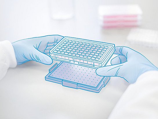
Bioprinting 96, 384 & 1536 well

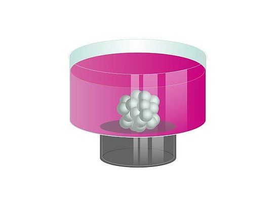
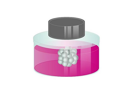
With bioprinting magnetised cells are printed into spheroids by placing a cell-repellent plate atop a drive of magnets, where a single magnet below each well utilises mild magnetic forces to induce aggregation and print one spheroid at the bottom of each well within 15 minutes to a few hours.

Levitation 6 & 24 well

By levitating cells from the bottom of a cell-repellent plate, magnetic forces work as an invisible scaffold to gently and rapidly aggregate cells, forming larger spheroids at the air-liquid interface, induce cell-cell interactions and initiate ECM synthesis.

Bio Assay 96 & 384 well

In addition bioprinting with a spheroidal shape, magnetic printing of cells can also be patterned into a ring formation. For up to 72 hours immediately following bioprinting, the patterned structures will shrink/close as a function of cell migration, viability, cell-cell interaction, and/or proliferation.

Advantages of the magnetic 3D cell culture (m3D) technology

  • 3D in a 2D workflow
  • Reproducible spheroid formation
  • Scalable – 6 Well to 1536 Well
  • Performed on a flat surface optimal for high-resolution microscopy and HTS
  • Rapid 3D culture formation within 24 hours for most cell types
  • No specialized equipment or media
  • Easy media changes and co-culture of different cell types
  • Compatible with fluorescence microscopy, Western blotting, qRT-PCR, Flow Cytometry, viability assays, chemiluminescence, etc.
  • Ready for automation
  • Biocompatible nanoparticles to magnetize cells

MAGNETISATION WITH NANOSHUTTLE-PL

NanoShuttle-PL is a nanoparticle assembly (~50 nm) consisting of biocompatible components: gold, iron oxide, and Poly-L-Lysine (PLL). Although NanoShuttle-PL is not itself an FDA-approved product for use in humans, the constituent components are themselves biocompatible. The cells are magnetised by electrostatically attaching small amounts of NanoShuttle-PL non-specifically to cell membranes via PLL at a concentration of around 50 pg/cell. Magnetised cells will appear peppered with dark nanoparticles after incubation, giving a speckled appearance. A small magnetic force of 30 pN/cell is enough to levitate and assemble cells without causing any harm.

NANOSHUTTLE-PL - THE BIOCOMPATIBILITY

  • Will not affect proliferation, viability, metabolism, inflammatory or oxidative stress, phenotype and/ or other cell functions
  • Does not bind any specific receptors, works with all cell types
  • Will release from the cell over 7-8 days into the surrounding extracellular matrix
  • Does not cause any chromosomal abnormalities in cells and does not lead to genomic instability

Magnetic 3D cell culture is an easy tool to create native tissue environments in vitro. With magnetic 3D bioprinting, the magnetized cells can be easily manipulated with magnetic fields to form spheroids by placing a cell-repellent plate containing the magnetized cells in media atop a drive of magnets. One magnet below each well utilizes mild magnetic forces to induce cell aggregation and print one spheroid at the bottom of each well anywhere within 15 minutes to a few hours. Afterwards the spheroids can be cultured long-term without the use of magnetic force. This system overcomes the limitations of other platforms by enabling rapid formation of spheroids, reproducible and scalable in size for high-throughput formats (96, 384 and 1536 well) and without limitation to cell types.

The 3D printing method together with commercially available standardized biochemical assay methods to facilitate continuous assessment of cell viability and other functions, provides an ideal combination for high-throughput compound screening.

By magnetized spheroids, adding and removing solutions is made easy by the use of magnetic forces to hold down spheroids during aspiration, limiting spheroid loss. Spheroids can also be picked up and transferred between vessels using magnetic tools such as the MagPen. Magnetic forces can also be used to create co-cultures with fine spatial organization.

In contrast to magnetic bioprinting, in magnetic 3D levitation, the magnetized cells are levitated off the bottom by a magnet above the plate. By levitating cells off the plate bottom the magnetic forces work as an invisible scaffold that rapidly and gently aggregates cells and induces cell-cell interactions and ECM synthesis. The 3D culture is formed without any artificial substrate or specialized media or equipment and can be cultured long-term. The 3D culture is formed without any artificial substrate or specialized media or equipment and can be cultured long-term.

The gentle nature of magnetic levitation allows cultures to acquire macroscale morphology that mostly resembles its tissue of origin. 3D cell cultures can be analyzed using common biological research techniques, such as immunohistochemical analysis and western blotting.

The magnetic levitation has been successfully used to make 3D cultures with different cell types, including different cell lines, including stem cells and primary cells.

The current standards for compound screening are animal models; while representing human tissues of interest, these models are expensive, scarce, and carry ethical challenges. On the other end, 2D in vitro assays poorly mimic native cellular environments and thus human in vivo response, but offer high-throughput testing with ease. There is a demand for in vitro assays that are both predictive of human in vivo response and high-throughput.

As a result, we developed a viability assay, the BiO Assay. Based on magnetic 3D bioprinting, cells magnetized with NanoShuttle-PL (NS) are printed into spheroids and rings.

Immediately after printing, these structures will shrink/close, as a function of cell migration, viability, cell-cell interaction, and/or proliferation, and varies with dosage. Culture contraction is generally complete within 24 hours, and images are batch processed to rapidly yield toxicity data Moreover, as the assay is label-free, the remaining rings or spheroids are available for further experimentation (IHC, Western blot, genomics, etc.).

The BiO Assay can be used to track the culture contraction of both rings and spheroids representing different situations. For rings, closure of the ring can represent wound-healing, wherein cells are working to close the void in the middle of the ring. Additionally, rings can represent similarly shaped tissues, like blood vessels, where dilation and contraction can be assayed. For Spheroids, contraction is related to spheroid assembly, with the assay macroscopically measuring how well the cells are interacting and migrating to build a competent structure.

The BiO Assay combines 3D cell culture environments with high-throughput and high-content testing to effectively predict in vivo response in vitro.

MagPen - Your smart assistant for 3D cell culture transfer

The MagPen facilitates easy and fast transfer and collection of magnetised cell cultures without disrupting their microtissue architecture. Cells, magnetised and cultured by Magnetic 3D Cell Culture (M3D), can be transposed by a simple “pick up-and-drop”-step. Additionally to that, the MagPen can be used to create and organize co-cultures by combining different magnetised 3D cell cultures.
The MagPen is availible as single version and as Multi-MagPen in 24 and 96 well format for simultaneous transfer of various cell cultures in one step.

MagPen - The principle

  • Fast, easy and simultaneous transfer of multiple 3D cell cultures without pipetting
  • Complete media change by a simple “pick up-and-drop”- step
  • Simplified co-culturing of different cell types
  • Ideal for easy immunohistochemistry staining, blocking, and washing of spheroids

ThinCert® Cell Culture Inserts

The ThinCert® cell culture inserts are suitable for a wide range of applications including:

  • Transport, secretion and diffusion studies
  • Migrational experiments
  • Cytotoxicity testing
  • Co-cultures
  • Trans epithelial electric resistance (TEER) measurements
  • Primary cell culture

Migration/ Invasion assay

Cell migration plays a significant role in physiological and pathological processes during embryonic development. As well as wound healing, immune response, inflammation, and tumorigenesis. The filter assay is a standard in vitromodel used to study cell migration. It is performed in cell culture inserts with 8.0 μm pores of the ThinCert® membrane. Cell migration is supported and involved from the upper compartment towards a chemo-attractant source in the lower compartment.

Co-Culture

Co-culture involves the study of immune cell interactions. Additionally, it includes other diverse applications such as the stimulation of cell proliferation, the maintenance of cell differentiation, and the restoration of heterocellular functions in vitro (e.g. bloodbrain-barrier). With ThinCert® cell culture inserts, cells may be seeded in the upper and lower compartment. The 0.4 µm or 1.0 µm pores of the ThinCert® membrane allow the exchange of molecules between the two cell populations.

Transport Studies

Transport studies are among the most frequent applications of cell culture inserts. The goal is to reconstruct a functional epithelium from individual cells. Additionally, an active transport of substances from one compartment through the epithelium into the other compartment will be investigated. ThinCert® cell culture inserts with 0.4 µm pores and translucent membranes are recommended for transport assays.

Organotypic and air-lift-culture

In organotypic culture a tissue can be kept alive for prolonged periods. While in tissue reconstruction the tissue is de novo generated from single cells. Both procedures use cell culture inserts with 0.4 to 3.0 µm pores. The tissue growth in vitro at the air-liquid-interface without limitations from gas exchange. Furthermore, for some tissue types, the direct exposure of the cultivated cells to the surrounding atmosphere serves as an indispensable differentiation stimulus.

  • Hanging geometry
  • Improved cell adhesion through physical surface treatment
  • Simplified pipetting due to self-lift geometry
  • Enhanced pipetting access and gas exchange

For light or electron microscopic examinations, the membranes can be easily detached from the housing using a scalpel. Even when detached, the membrane remains flat without unrolling, greatly simplifying further steps. Due to its high chemical resistance to various solvents, the membrane is suitable for numerous cell fixation protocols. Due to their special suspension, ThinCert® cell culture inserts keep a distance to the well bottom at all times, which means that cells cultured there are always protected from damage. A safe minimum distance to the side walls is always maintained with the help of distance holders, which prevents capillary suction between the inner wall of the well and the outer wall of the insert. This means that mass transfer between the well and ThinCert® takes place exclusively via the porous membrane. ThinCert® cell culture inserts sit eccentrically in the wells and deflect upwards when a pipette is inserted. When the pipette is removed again, the ThinCert® automatically resume their original position. This feature is called self-lift geometry.

  • Small pore sizes (0.4 and 1 μm in diameter): For co-cultivation, transportation, secretion, and diffusion studies
  • Larger pore sizes (3 and 8 μm in diameter): For migration and invasion studies
  • Transparent membranes (in general low pore density): Suitable for light and electron microscopy. Not suited for all applications requiring high diffusion rates such as transport studies
  • Translucent membranes (in general high pore density): Suitable for electron microscopy, fluorescence microscopy, confocal microscopy, TEER and transport studies

Material:

ThinCert® cell culture inserts are produced from high-grade clear polystyrene housings, and polyethylene terephthalate (PET) capillary pore membranes. Both materials, polystyrene and PET, are USP class VI certified and cell culture compatible. The coupling between the housing and the membrane is achieved using an automated process. This produces an extremely strong and robust seal without compromising or weakening the membrane in any way.

The membranes undergo a physical surface treatment to optimise cellular adherence and growth characteristics. All the capillary pores in a membrane exhibit a high degree of uniformity in diameter. This uniformity ensures reliable and consistent exchange rates between the two compartments and thus provides reproducibility when conducting multiple experiments.

Kits:

The cell culture inserts are pre-packed together with the requisite number of plates. The automated production process includes double optical control of each insert produced, ensuring that any biological contamination is avoided. The sterility of the single blisterpacked inserts and multiwell plates is ensured by irradiation.

CELLSTAR® Cell-Repellent Surface

In contrast to standard tissue culture surfaces which are optimised to enhance conditions for cell attachment, the cell-repellent surface has been developed to effectively prevent cell attachment. CELLSTAR® cell culture vessels with a cell-repellent surface reliably prevent cell attachment in suspension cultures of semiadherent and adherent cell lines where standard hydrophobic surfaces generally used for suspension culture are insufficient.

For formation of spheroids, stem cell aggregates and self-assembled spherical clusters used as 3D cell culture models, the cell-cell interaction must dominate over the interaction between the cells and the culture surface of containment.

Therefore CELLSTAR® cell culture vessels with cell-repellent surface effectively prevent cell adherence and promote the spontaneous formation of three-dimensional spheroids by gravitation: a single spheroid per well in round bottom microplates or multiple spheroids in flat bottom plates, dishes and flasks.

Long-term incubations of hydrogel cultures are frequently performed as an approach to mimic a 3D environment. When standard tissue culture vessels are used in this approach, some cells tend to migrate out of the hydrogel, forming a 2D subculture on the vessel surface. Analysis of such a cell population will therefore result in mixed data from both 2D and 3D cell cultures. CELLSTAR® cell culture vessels with a cell-repellent surface can be used for hydrogel cultures to effectively suppress the formation of 2D subcultures.

Applications & Cell Lines

What 3D platform is best for your application?

Stem Cell Lines

Cancer Cell Lines

Primary Cell Lines

Other Cell Lines

Pre-adipocyte stem cell

KPC pancreatic ductal adenocarcinoma


Pancreatic ß-cells (EndoC-ßH3)

Neural stem cells

LN229 – Glioblastoma cells

Primary Glioblastoma cells

Human Astrocyte

Neural crest-derived mesenchymal stem cell

A549 — Lung epithelial adecarcenoma

Valvular interstitial cells (VICs)

Bend (brain endothelial)

Mesenchymal stem cell

HepG2 — Human liver carcinoma

Human lung primary cells:

  • epithelial
  • endothelial
  • fibroblasts
  • smooth muscle

3T3 fibroblasts

Dental pulp stem cell

PC3 — Human prostate cancer

Valvular endothelial cells (VECs)

Adipocyte


H-4-II-E — Rat hepatoma, liver

Aortic valve co-cultures (AVCCs)

Huvec - Human umbilical endothelial cells


MDA-231 - Human breast cancer

Primary mouse heart cells

HPF - Human pulmonary fibroblast


LNCaP - Prostate cancer cell line

Human Primary vascular smooth muscle

SMC - Tracheal smooth muscle cell


Ovarian cancer cells

Primary Miomytrial Smooth muscle

HEK293 - Human embryonic kidney


Panc-1 - Pancreatic cancer cell

Primary human hepatocytes

MCF-10A - Breast epithelial cell line


Cancer associated fibroblasts

Primary pancreatic cancer cells

Fibroblast


Triple negative inflammatory breast cancer

Primary tissue from PDX

Chondrocytes


Caki-1 – Human renal cancer cell line

Primary fibroblasts

T-cells


Osteosarcoma

Keratinocytes

A10 - Rat vascular smooth muscle


HCT116 - colon cancer cell line


DU145


Murine embryonic fibroblasts (iMEF)


LOVO cells


DT66066 cells


Spinal cord cells


Endothelial cell


Peripheral blood mononuclear cells (PBMCs)


Monocytes

Publications

Year

Author / Title / Link

Product

Application Area

Cell Type

2022

Chen, G.; Liu, W.; Yan, B.: Breast Cancer MCF-7 Cell Spheroid Culture for Drug Discovery and Development

Cell-Repellent

Breast Cancer

MCF-7 cells (Human Breast Cancer)

2022

Neebe et al. Small integral membrane protein 10 like 1 downregulation enhances differentiation of adipose progenitor cells Biochemical and Biophysical Research Communications

Cell-Repellent

Cancer / Lipoma Formation

Lipoma Cells

2022

Seitlinger et al. Vascularization of Patient-Derived Tumoroid from Non-Small-Cell Lung Cancer and Its Microenvironment

ThinCert®

Cancer

Human Pulmonary Fibroblasts,

Human Umbilical Vein Endothelial Cells,

Human Mesenchymal Stem Cells,

Lung Tumor Cells 

2021

Batista et al. Three-Dimensional Adipocyte Culture as a Model to Study Cachexia-Induced White Adipose Tissue Remodeling

m3D

Cancer Research

Stem Cells

Adipose Tissue,

Sem Cell Adipocyte

2021

Jaromi et al. KRAS and EGFR Mutations Differentially Alter ABC Drug Transporter Expression in Cisplatin-Resistant Non-Small Cell Lung Cancer

m3D

Cancer Resaerch

A549 (KRAS),

PC9 (EGFR)

2020

Kiss et al. Cisplatin treatment induced interleukin 6 and 8 production alters lung adenocarcinoma cell migration in an oncogenic mutation dependent manner  

m3D

Cancer Research

Migration/Invasion Assay

Wound Healing

Primary Lung Cancer,

549 and PC-9 Human Lung Adenocarcinoma

2020

Souza-Araújo et al. Three-Dimensional Cell Culture Based on Magnetic Fields to Assemble Low-Grade Ovarian Carcinoma Cell Aggregates Containing Lymphocytes

m3D

Cancer Research

Patient Cells,

Ovarian Carcinoma,

Lymphocytes

2019

Leenhardt et al. Ultrasound-induced cavitation enhances the efficacy of Chemotherapy in a 3D Model of Pancreatic Ductal Adenocarcinoma with its microenvironment

m3D

Cancer Research

Murine Embryonic Fibroblasts (iMEF),

KPC Pancreatic Ductal Adenocarcinoma (PDA),

T66066 Cells

2019

Mejía-Cruz et al. Generation of Organotypic Multicellular Spheres by Magnetic Levitation: Model for the Study of Human Hematopoietic Stem Cells Microenvironment

m3D

Cancer Research

Stem Cells

Human Hematopoietic Stem Cells,

Human Bone Marrow-Mesenchymal Stem Cells,

Umbilical Cord Blood-Hematopoietic Stem Cells

2018

Degadi et al. Klotho inhibits EGF-induced cell migration in Caki-1 cells through inactivation of EGFR and p38 MAPK signaling pathways

m3D

Cancer Research

Wound Healing

Caki-1

2018

Eckhardt et al. Clinically relevant inflammatory breast cancer patient-derived xenograft-derived ex vivo model for evaluation of tumor-specific therapies

m3D

Cancer Research

Screening / Toxicity

Triple Negative Inflamatory Breast Cancer

2018

Hou et al. Advanced Development of Primary Pancreatic Organic Tumor Models for High-Throughput Phenotypic Drug Screening

m3D

Cancer Research

Screening / Toxicity

Triple Negative Inflamatory Breast Cancer

2017

Noel et al. Preparation and Metabolic Assay of 3-dimensional Spheroid Co-cultures of Pancreatic Cancer Cells and Fibroblasts

m3D

Cancer Research,

Co-Culture

Panc-1 - Pancreatic Cancer Cell

Fibroblast

2016

Hau et al. Dose enhancement and cytotoxicity of gold nanoparticles in colon cancer cells when irradiated with kilo‐ and mega‐voltage radiation

m3D

Cancer Research

Migration/Invasion Assay

LOVO cells,

Colon adenocarcinoma

2016

Pan et al. miR-509-3p is clinically significant and strongly attenuates cellular migration and multi-cellular spheroids in ovarian cancer

m3D

Cancer Research

Wound Healing

Ovarian ccancer cells

2014

Jaganathan et al. Three-dimensional in vitro co-culture model of breast tumor using magnetic levitation

m3D

Cancer Research

Tissue Engineering / Reconstruction

Co-Culture

MDA-231,

Fibroblast

2013

Becker, J. L.; Souza, G. R.: Using space-based investigations to inform cancer research on Earth

m3D

Cancer Research

Glioblastoma

2010

Becker, J. L.; Souza, G. R.: Invasive glioblastoma cells acquire stemness and increased Akt activation

m3D

Cancer Research

Stem Cells

LN229 - Glioblastoma,

Normal Human Astrocyte,

Primary Glioblastoma

2010

Glauco et al. Three-dimensional tissue culture based on magnetic cell levitation

m3D

Cancer Research

Stem Cells

Co-Culture

LN229 - Glioblastoma,

Normal Human Astrocyte,

Neural Stem Cell

Year

Author / Title / Link

Product

Application Area

Cell Type

2022

Baarsma et al. Epithelial 3D-spheroids as a tool to study air pollutant-induced lung pathology

m3D

Screening/Toxicity

Human Bronchial Epithelial (BEAS-2B)

2022

Cromwell et al. Multifunctional profiling of triple-negative breast cancer patient-derived tumoroids for disease modeling

m3D

Screening/Toxicity

Cancer Research

Microfluids

Patient-Derived Tumor Explant,

TU-BcX-4IC,

Breast Cancer,

PDX Organoids (PDX-O) 

2022

Fernandez-Vega et al. Lead Identification using 3D Models of Pancreatic Cancer

m3D

Screening/Toxicity

HTS

Patient Samples

Pancreatic Cancer Cells

2021

Rodboon et al. Development of high-throughput lacrimal gland organoid platforms for drug discovery in dry eye disease

m3D

Screening/Toxicity

Stem Cells

Myoepithelial Cells (MEC)

Lacrimal Gland Ctem Cells

2021

Trindade Caleffi et al. Magnetic 3D cell culture: State of the art and current advances

m3D

Screening/Toxicity

Cardiomyocytes,

Bronchial and Pancreatic Cells

2020

Burnham et al. A Scalable Approach Reveals Functional Responses of iPSC Cardiomyocyte 3D Spheroids

m3D

Screening/Toxicity

Electrophysiology

Cardiomyocytes and Fibroblast

Co-Culture

2019

Baillargeon et al. Automating a Magnetic 3D Spheroid Model Technology for High-Throughput Screening

m3D

Screening/Toxicity

Cancer Research

Pancreatic Cancer Cells

2018

Souza et al. Comparative Assay of 2D and 3D Cell Culture Models: Proliferation, Gene Expression and Anticancer Drug Response

m3D

Screening/Toxicity

Cancer Research

Prostate Cancer Cell Lines: PC-3,

LNCaP and DU145

2017

Desai, P. K.; Tseng, H.; Souza, G. R.: Assembly of hepatocyte spheroids using magnetic 3D cell culture for CYP450 inhibition/induction

m3D

Screening/Toxicity

Primay Human Hepatocytes

2017

Souza et al. Magnetically bioprinted human myometrial 3D cell rings as a model for uterine contractility

m3D

Screening/Toxicity

Primary Miomytrial Smooth Muscle

2016

Tseng et al. A high-throughput in vitro ring assay for vasoactivity using magnetic 3D bioprinting

m3D

Screening/Toxicity

Wound Healing

A10 - Rat Vascular Smooth Muscle,

Human Primary Vascular Smooth Muscle

2015

Tseng et al. A spheroid toxicity assay using magnetic 3D bioprinting and real-time mobile device-based imaging

m3D

Screening/Toxicity

3T3 Fibroblast

2013

Haisler et al. Three-dimensional cell culturing by magnetic levitation

m3D

Screening

Tissue Engineering / Reconstruction

Cancer Research

HepG2 - Human Liver Carcinoma,

A549 - Lung Epithelial Adecarcenoma,

PC3 - Human Prostate Cancer,

LN229 - Glioblastoma,

Huvec - Human Umbelical Endothelial Cells,

H-4-II-E - Rat Hepatoma, Liver,

T3T - Mouse fFbroblast,

HPF - Human Pulmonary Fibroblast,

SMC - Tracheal Smooth Muscle,

MDA-231 - Human Breast Cancer(a),

HEK293 - Human Embrionic Kidney,

MCF-10A - Breast Epithelial Cell Line,

LNCaP - Prostate Cancer Cell Line

Year

Author / Title / Link

Product

Application Area

Cell Type

2022

Baarsma et al. Epithelial 3D-spheroids as a tool to study air pollutant-induced lung pathology

Cell-Repellent

Lung pathology

Human Bronchial Epithelial (BEAS-2B)

2022

Castillo et al. Human Air-Liquid-Interface Organotypic Airway Cultures Express Significantly More ACE2 Receptor Protein and Are More Susceptible to HCoV-NL63 Infection than Monolayer Cultures of Primary Respiratory Epithelial Cells

ThinCert®

COVID

Primary Human Respiratory Epithelial Cells

2022

Chansaenroj et al. Magnetic bioassembly platforms towards the generation of extracellular vesicles from human salivary gland functional organoids for epithelial repair

m3D

Tissue Engineering / Reconstruction

Stem Cells

Human Dental Pulp Stem Cells (hDPSC),

Salivary Gland

2022

Tan, C. T., Leo, Z. Y., Lim, C. Y.: Generation and integration of hair follicle-primed spheroids in bioengineered skin constructs

ThinCert®

Skin research

N/TERT-1 Keratinocytes,

Human Primary Adult Keratinocytes,

Primary Neonatal Keratinocytes,

Human Hair Follicle DP Cells 

2021

Bing, et al. Human organoid biofilm model for assessing antibiofilm activity of novel agents

ThinCert®

Skin Model / Biofilm

N/TERT Keratinocytes

2019

Bowser, D. A.; Moore, M. J.: Biofabrication of neural microphysiological systems using magnetic spheroid bioprinting

m3D

Tissue Engineering / Reconstruction

Spinal Cord Cells

2018

Tseng et al. Three-Dimensional Magnetic Levitation Culture System Simulating White Adipose Tissue

m3D

Tissue Engineering / Reconstruction

Endothelial Cell,

Pre-Adipocyte Stem Cell

2016

Hogan et al. Assembly of a functional 3D primary cardiac construct using magnetic levitation

m3D

Tissue Engineering / Reconstruction

Primary Mouse Heart Cells,

Fibroblast,

Endothelial Cells

2016

Lin et al. Nanoparticle improved stem cell therapy for erectile dysfunction in a rat model of cavernous nerve injury

m3D

Tissue Engineering / Reconstruction

Stem Cells

Mesenchymal Stem Cell

2013

Timm et al. A high-throughput three-dimensional cell migration assay for toxicity screening with mobile device-based macroscopic image analysis

m3D

Wound Healing

Screening/Toxicity

HEK293 - Kidney,

Primary Tracheal Smooth Muscle

2013

Tseng et al. A three-dimensional co-culture model of the aortic valve using magnetic levitation

m3D

Tissue Engineering / Reconstruction 

Co-Culture

Valvular Interstitial Cells (VICs),

Valvular Endothelial Cells (VECs),

Aortic Valve Co-Cultures (AVCCs)

2013

Tseng et al. Assembly of a three-dimensional multitype bronchiole coculture model using magnetic levitation

m3D

Tissue Engineering / Reconstruction 

Primary Human Lung Cells: Epithelial, Endothelial, Fibroblast, Smooth Muscle

Year

Author / Title / Link

Product

Application Area

Cell Type

2021

Avelino et al. Mass spectrometry-based proteomics of 3D cell culture: A useful tool to validate culture of spheroids and organoids

m3D

Stem Cells

Tissue Engineering / Reconstruction

3T3,

Pre-Adipocyte Stem Cells,

Adipocytes

2019

Ferreira et al. A magnetic three‐dimensional levitated primary cell culture system for the development of secretory salivary gland‐like organoids

m3D

Stem Cells

Salivary Gland Derived Cells

2018

Adine et al. Engineering innervated secretory epithelial organoids by magnetic three-dimensional bioprinting for stimulating epithelial growth in salivary glands

m3D

Stem Cells

Tissue Engineering / Reconstruction

Neural Crest-Derived Mesenchymal Stem Cell,

Dental Pulp Stem Cell

2013

Daquinag, A. C., Souza, G. R.; Kolonin, M. G.: Adipose tissue engineering in three-dimensional levitation tissue culture system based on magnetic nanoparticles

m3D

Stem Cells

Tissue Engineering / Reconstruction

Primary Stroma,

3T3,

Bend (Brain Endothelial)  

Year

Author / Title / Link

Product

Application Area

Cell Type

2021

Vu et al. Scaffold-free 3D cell culture of primary skin fibroblasts induces profound changes of the matrisome

m3D

Tissue Engineering / Reconstruction

Primary Skin Fibroblasts

2019

Antonino et al. Three-dimensional levitation culture improves in-vitro growth of secondary follicles in bovine model

m3D

-

Bovine Secondary Follicles,

Oocyte

2019

Klinder et al. Comparison of different cell culture plates for the enrichment of non-adherent human mononuclear cells

Cell-Repellent

-

Peripheral Blood Mononuclear Cells (PBMCs),

Monocytes

2019

Nagaraju et al. Myofibroblast modulation of cardiac myocyte structure and function

ThinCert®

Myocardial Infarction

Cardiomyocytes,

Fibroblasts

2019

Urbanczyk et al. Controlled Heterotypic Pseudo-Islet Assembly of Human b-Cells and Human Umbilical Vein Endothelial Cells Using Magnetic Levitation

m3D

Co-Culture

Tissue Engineering / Reconstruction

Pancreatic ß-Cells (EndoC-ßH3),

Human Umbilical Vein Endothelia Cells (HUVECs)

2013

Castro-Chavez et al. Effect of lyso-phosphatidylcholine and Schnurri-3 on osteogenic transdifferentiation of vascular smooth muscle cells to calcifying vascular cells in 3D culture

m3D

-

Human Umbilical Vein Endothelia Cells (HUVECs)

Downloads & Media

Magnetic 3D Cell Culture - Tools to Bridge the Gap Between 2D and 3D

Presenter: Gluaco R. Souza

Year: 2020, Language: English

Learning objectives:

  • General technique of magnetic 3D cell culture and magnetic 3D bioprinting
  • Key advantages of 3D cell culture over other techniques
  • Broad applications of 3D cell culture
  • Applications in immunobiology
  • Comparison between magnetic 3D cell culture and in vivo results in collaboration with MD Anderson Cancer Center

Automated spheroid production by magnetic cell assembly for reproducible HTS applications

Presenter: Glauco R. Souza (Greiner Bio-One) & Michael Nosswitz (Tecan Switzerland)
Year: 2023, Language: English

Learning objectives

  • Learn how to utilize magnetic 3D bioprinting to enable automation and optimal imaging conditions
  • Discover how to automate spheroid production to reduce uniformity variations
  • Consider how to automatically monitor QC for spheroid formation and production
  • Find out how to simplify and automate drug dosing for synergy experiments

Year

Title

Conference

2019

Three-dimensional cytotoxicity assay using magnetic 3D bioprinting for measuring CAR T cell function in heterogeneous solid tumor microenvironments

American Association of Cancer Research Annual Meeting, Atlanta, GA

2018

Automated, Image-Based T Cell Mediated Cytotoxicity Assessments using 2D and 3D Target Cell Models

SLAS 2018

2018

3D Cultures of iPSC-derived Human Motor Neurons & Tracheal Smooth Muscle Cells in HTS Format Using Magnetic 3D Bioprinting

Society for Neuroscience Annual Meeting, San Diego, CA

2018

A PDX-DerivedEx-Vivo Tumor Tissue Array Platform Utilizing Magnetic 3D Bioprinting for The Identification of Tumor-Specific Therapies

American Association of Cancer Research Annual Meeting, Chicago, IL

2018

Validation of Magnetic 3D Spheroid Bioprinting in Combination with a BlueWasher

Society of Lab Automation and Screening Annual Conference and Exhibition, San Diego, CA

2016

Development of spheroids derived from tumor biopsies and patient-derived xenografts using magnetic 3D bioprinting

American Association of Cancer Research Annual Meeting, New Orleans, LA

2016

High-throughput spheroid formation for compound screening using magnetic 3D bioprinting

Dechema 3D Cell Culture, Freiburg, Germany

2016

Improvement of Human iPS Cell-Derived Hepatocyte Functionality Using 3D Culture System


2016

Magnetically 3D bioprinted hepatocyte spheroids for in vitro metabolic studies

Society of Toxicology Annual Meeting, New Orleans, LA

2016

High-throughput spheroid formation for compound screening using magnetic 3D bioprinting

Society of Lab Automation and Screening Annual Conference and Exhibition, San Diego, CA

2015

Somatic mutation detection from liquid biopsy-derived cellular aggregates formed by magnetic 3D bioprinting

AACR-NCI-EORTC International Conference on Molecular Targets and Cancer Therapeutics, Boston, MA

2015

High-throughput functional toxicity screening with iPS-cardiomyocyte and hepatocyte spheroids by magnetic 3D bioprinting

Cellular Dynamics iForum, Chicago, IL

2015

High-throughput spheroid formation in a 384-well format using magnetic 3D bioprinting

American Association of Cancer Research Annual Meeting, Philadelphia, PA

2015

High-throughput spheroid printing and toxicity testing using magnetic 3D bioprinting

Society of Lab Automation and Screening Annual Conference and Exhibition, Washington, DC

2014

Magnetic 3D Bioprinting: A novel high-throughput and high-content assay for toxicity screening

European Society of Toxicology In Vitro International Conference, Egmond aan Zee, The Netherlands

2014

A novel vascular “ring” assay for smooth muscle contractility using magnetic 3D bioprinting

Arteriosclerosis, Thrombosis, and Vascular Biology Scientific Sessions, Toronto, ON

2014

Magnetic 3D Bioprinting: A novel high-throughput and high-content assay for toxicity screening

Society of Toxicology Annual Meeting, Phoenix, AZ

2013

A high-throughput three-dimensional magnetically printed cellular assay (BiO Assay) for toxicity screening for breast cancer applications

San Antonio Breast Cancer Symposium, San Antonio, TX

2013

A high-throughput three-dimensional cell migration assay for toxicity screening using magnetic levitation with mobile device-based macroscopic image capture

American Association of Pharmaceutical Scientists Annual Meeting and Exhibition, San Antonio, TX

FAQs

Organoids and spheroids are both 3D cell culture models, but they differ in their complexity and the types of cells they are composed of. Spheroids are typically simpler, consisting of one or two cell types, and are often used for drug testing and cancer research. On the other hand, organoids are more complex and contain several types of cells that organize themselves to mimic the architecture and functionality of an organ. Organoids can be derived from stem cells or organ progenitors and are used for studying organ development, disease modeling, and personalized medicine.

The choice between using organoids or spheroids depends on the research question. Spheroids are often used in cancer research for studying tumor growth, invasion, and drug response. They are also used in toxicity testing. Organoids are used when a more complex and organ-like structure is needed, such as in studies of organ development, disease modeling, and drug testing in a more organ-relevant context.

Spheroids and organoids are useful in drug screening because they mimic the 3D structure of tissues and organs more accurately than 2D cell cultures. This allows for a more accurate prediction of how drugs will behave in the body. They can be used to test drug efficacy and toxicity before moving on to animal models, reducing the cost and time of drug development.

  • Magnetic 3D Cell Culture by Levitation and Bioprinting:
    This method uses gentle magnetic fields to levitate cells or bioprint them in 3D structures. The cells are first magnetized using a nanoparticle assembly called NanoShuttle-PL, which adheres to the cell membrane. The cells can then be manipulated using magnetic fields to form 3D structures. This method is easy to use, efficient, and reproducible, making it a popular choice for generating spheroids, especially when using flat-bottom plates. More information about the biocompatibility of NanoShuttle can be found in this whitepaper
     
  • Scaffold-free Techniques:
    These techniques rely on the natural ability of cells to self-assemble into 3D structures. One common method is to use CELLSTAR® cell-repellent plates with flat or round bottom plates. These plates have a low-attachment surface that prevents cell adhesion, promoting cells to interact with each other and form 3D structures. The round bottom plates facilitate cell aggregation.
     
  • Scaffold-based techniques:
    These techniques use a supporting material, or scaffold, to provide a 3D structure for the cells to grow on. The scaffold can be made from a variety of materials, including natural polymers like collagen or synthetic materials like polyethylene glycol. Hydrogels, which are water-swollen networks of polymer chains, are a common type of scaffold used in 3D cell culture. They can mimic the extracellular matrix and allow for the diffusion of nutrients and growth factors.
     
  • Organoid culture:
    Organoids are self-organizing 3D structures that are derived from stem cells or organ progenitors and can mimic the architecture and functionality of specific organs. They are grown in a supportive matrix, often a hydrogel, and are guided to differentiate into the desired cell types through the use of specific growth factors and signaling molecules.
     
  • Microfluidic systems:
    These systems use microscale channels to control the flow of cell culture medium and allow for the precise control of the cell culture environment. They can be used to generate 3D cell cultures and are particularly useful for creating organ-on-a-chip models.
     
  • Bioprinting:
    This is a relatively new technique that uses 3D printing technology to create complex 3D structures of cells and biomaterials. It allows for a high degree of control over the spatial distribution of cells and can be used to create tissue-like structures.

The NanoShuttle-PL is an essential part of the magnetic 3D cell culture technology. It is responsible for the magnetization of the cells. Once magnetized, the cells can be aggregated by magnetic forces and these cells interact and self-assemble into a culture that recreates in vivo environments. In addition, the magnetic feature can be used to hold the 3D cell culture in place while processing. A common concern is the potential for toxic or other adverse effects of NanoShuttle-PL. Various research with this platform from our lab and within independent studies of our users has shown so far no effect of NanoShuttle-PL on cell health or function. This application note will discuss in further detail the results demonstrating the biocompatibility of NanoShuttle-PL source.

Magnetic 3D cell culture and traditional methods like round bottom or hanging drop both enable the formation of 3D cell cultures, but they differ significantly in their approach and capabilities. Magnetic 3D cell culture, which uses magnetic fields to levitate and organize cells into 3D structures, offers advantages in terms of ease of manipulation, co-culturing of cells, and compatibility with automation. It allows for precise control over cell organization, facilitates tasks such as changing media and staining, and is well-suited for high-throughput applications. In contrast, traditional methods rely on gravity to form cell aggregates into 3D structures. While these methods are more straightforward and less expensive, they offer less control over cell organization, are less compatible with automation, and do not allow for easy changing of media and staining and other downstream manipulation steps.

The membrane thickness is identical for 6, 12 or 24 well types:


                                  OPTICAL                                                                       PRODUCT
                               MEMBRANE                     MEMBRANE                    EXAMPLE
Ø PORE               PROPERTIES                  THICKNESS                      ITEM NO.

0.4 µm                 transparent                      22 +/- 3 µm                        662641

1.0 µm                  transparent                      22 +/- 3 µm                        662610   

3.0 µm                 transparent                      20 +/- 3 µm                        662630

0.4 µm                 translucent                      22 +/- 3 µm                        662640

3.0 µm                 translucent                      20 +/- 3 µm                        662631

8.0 µm                 translucent                       15 +/- 3 µm                        662638

The membrane is TC treated from both sides and it is hydrophilic.

The pores are not parallel to one another but are angled to avoid the formation of multiple pores and hence the formation of larger pore sizes due to combination of individual pores.

  • 6 Well: Item No. 657160 CELL CULTURE MULTIWELL PLATE
  • 12 Well: Item No. 665180 CELL CULTURE MULTIWELL PLATE
  • 24 Well: Item No. 662160 CELL CULTURE MULTIWELL PLATE

Cell Repellent plates with round bottom are offered in a 96- and 384-well format, F-bottom plates with 6 to 1536 wells. With V-bottom there is a 96-well format.

Magnetic 3D is designed in combination with Cell Repellent F-bottom plates. This allows the formation of spheroids and imaging applications in the same plate.

The so-called μClear® bottom is a transparent thin film bottom. It is ideal for microscopic applications.

Events

COMING SOON

Explore the world of 3D cell culture

Contact us!
Please contact us if you have any questions regarding our innovative 3D Cell Culture products!

Please contact us if you have any questions regarding our innovative 3D Cell Culture products!

Glauco Souza, PH.D.

Director of Global Business Development & Innovation 3D Cell Culture

Sign-up to our newsletter or request more information here
Register now and you will receive our regular Greiner Bio-One News on current topics to stay up-to-date.

We redirected you to your country page. To go back to the previous page, please click the button.

You are not viewing your country page. To switch to your country, please click the button.